Copyright
©2007 Baishideng Publishing Group Co.
World J Gastroenterol. Mar 14, 2007; 13(10): 1477-1486
Published online Mar 14, 2007. doi: 10.3748/wjg.v13.i10.1477
Published online Mar 14, 2007. doi: 10.3748/wjg.v13.i10.1477
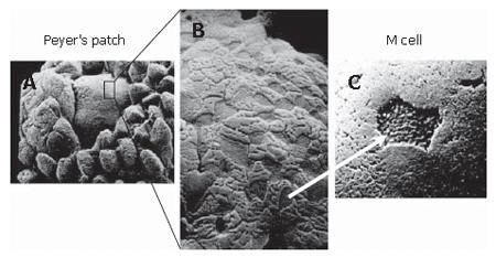
Figure 1 Ultrastructure of the Peyer’s patches and FAE (Adapted by permission from Macmillan Publishers Ltd: Nature Reviews Immunology[32], copyright 2003).
A: At low magnification, the dome shape of the Peyer’s patch protrudes between villi into the lumen of the intestine; B: At higher magnification, M cells can be seen as epithelial cells with surface microfolds rather than the microvilli that are seen on the surrounding conventional enterocytes; C: Antigen is taken up preferentially through M cells.
- Citation: Miller H, Zhang J, KuoLee R, Patel GB, Chen W. Intestinal M cells: The fallible sentinels? World J Gastroenterol 2007; 13(10): 1477-1486
- URL: https://www.wjgnet.com/1007-9327/full/v13/i10/1477.htm
- DOI: https://dx.doi.org/10.3748/wjg.v13.i10.1477


