Copyright
©2007 Baishideng Publishing Group Co.
World J Gastroenterol. Jan 7, 2007; 13(1): 48-64
Published online Jan 7, 2007. doi: 10.3748/wjg.v13.i1.48
Published online Jan 7, 2007. doi: 10.3748/wjg.v13.i1.48
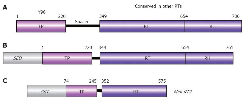
Figure 5 Domain structure of P protein.
A: Authentic DHBV P protein. Numbers are aa positions for DHBV P protein. The priming Tyr residue Y96 is indicated; B: Typical recombinant P protein construct. For solubility, a heterologous solubility enhancing domain (SED) such as NusA, GrpE, or GST is required, and a short stretch of C terminal aa must be removed. Deletion of the spacer has no negative effects on in vitro activity; C: Mini-RT2. This heavily truncated recombinant DHBV P protein requires mild detergent, but no chaperones for priming activity.
- Citation: Beck J, Nassal M. Hepatitis B virus replication. World J Gastroenterol 2007; 13(1): 48-64
- URL: https://www.wjgnet.com/1007-9327/full/v13/i1/48.htm
- DOI: https://dx.doi.org/10.3748/wjg.v13.i1.48


