Copyright
©2006 Baishideng Publishing Group Co.
World J Gastroenterol. Mar 7, 2006; 12(9): 1472-1475
Published online Mar 7, 2006. doi: 10.3748/wjg.v12.i9.1472
Published online Mar 7, 2006. doi: 10.3748/wjg.v12.i9.1472
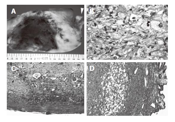
Figure 4 Resected specimen and histological findings.
A: Cutting section of the tumor; B: The main tumor; C: The edge of the tumor(hepatic side); D: The posterior wall of the gallbladder. The cutting section of the tumor appeared whitish solid and included parts of hemorrhage and necrosis and attached to the gallbladder firmly (A) (arrowhead: gallbladder). On histological examination, the tumor was mainly composed of spindle cells with cellular pleomorphism, and included lipoblasts (B). The tumor had a capsule (C), but the capsule annihilated a border of the gallbladder, where these tumor cells were detected in the muscle layer of the gallbladder (D).
- Citation: Hamada T, Yamagiwa K, Okanami Y, Fujii K, Nakamura I, Mizuno S, Yokoi H, Isaji S, Uemoto S. Primary liposarcoma of gallbladder diagnosed by preoperative imagings: A case report and review of literature. World J Gastroenterol 2006; 12(9): 1472-1475
- URL: https://www.wjgnet.com/1007-9327/full/v12/i9/1472.htm
- DOI: https://dx.doi.org/10.3748/wjg.v12.i9.1472


