Copyright
©2006 Baishideng Publishing Group Co.
World J Gastroenterol. Mar 7, 2006; 12(9): 1472-1475
Published online Mar 7, 2006. doi: 10.3748/wjg.v12.i9.1472
Published online Mar 7, 2006. doi: 10.3748/wjg.v12.i9.1472
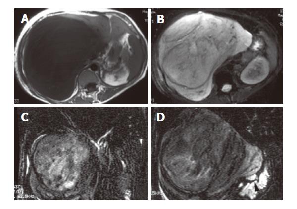
Figure 2 Preoperative MRI images.
A: T1-weighted;B:fat-supressed T2-weighted;B and C: MR Cholangiography. The tumor appeared hypointense on T1-weighted images (A) and hyperintense on fat-suppressed T2-weighted images (B). MR cholangiography revealed that the tumor compressed the right lobe of the liver to the cranial side (C) and gallbladder to the medial-caudal side(arrow head:gallbladder) (D).
- Citation: Hamada T, Yamagiwa K, Okanami Y, Fujii K, Nakamura I, Mizuno S, Yokoi H, Isaji S, Uemoto S. Primary liposarcoma of gallbladder diagnosed by preoperative imagings: A case report and review of literature. World J Gastroenterol 2006; 12(9): 1472-1475
- URL: https://www.wjgnet.com/1007-9327/full/v12/i9/1472.htm
- DOI: https://dx.doi.org/10.3748/wjg.v12.i9.1472


