Copyright
©2006 Baishideng Publishing Group Co.
World J Gastroenterol. Feb 28, 2006; 12(8): 1211-1218
Published online Feb 28, 2006. doi: 10.3748/wjg.v12.i8.1211
Published online Feb 28, 2006. doi: 10.3748/wjg.v12.i8.1211
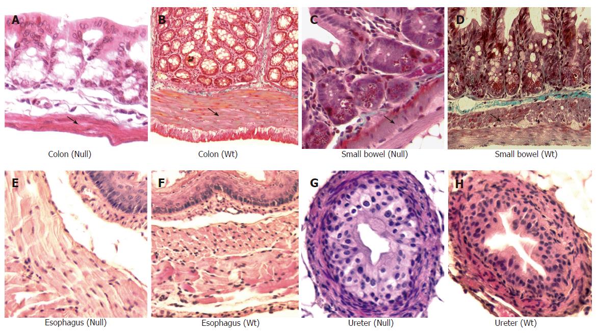
Figure 5 Masson trichrome staining (x 20) of small and large bowel from Smad3 mice.
Significant reduction of muscular layer of descending colon of Smad3 null (A) is observed compared to colon from wild-type (WT) mice (B), reduction of muscle layer in cross sections of the proximal small bowel from Smad3 null (C) as compared to wild-type mice (D). Haematoxylin and eosin staining (x 20) of ureter and esophagus of Smad3 mice. Cross section of esophagus from Smad3 null (E) and wild-type mice (F) shows no differences in muscle layer. Cross section of ureter from Smad3 null (G) and wild-type mice (H) shows no differences in muscle layers.
- Citation: Zanninelli G, Vetuschi A, Sferra R, D’Angelo A, Fratticci A, Continenza MA, Chiaramonte M, Gaudio E, Caprilli R, Latella G. Smad3 knock-out mice as a useful model to study intestinal fibrogenesis. World J Gastroenterol 2006; 12(8): 1211-1218
- URL: https://www.wjgnet.com/1007-9327/full/v12/i8/1211.htm
- DOI: https://dx.doi.org/10.3748/wjg.v12.i8.1211


