Copyright
©2006 Baishideng Publishing Group Co.
World J Gastroenterol. Feb 21, 2006; 12(7): 1078-1085
Published online Feb 21, 2006. doi: 10.3748/wjg.v12.i7.1078
Published online Feb 21, 2006. doi: 10.3748/wjg.v12.i7.1078
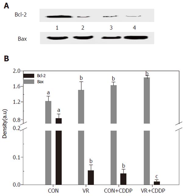
Figure 6 Expression of apoptosis regulatory proteins (Bcl-2 and Bax) in the intestinal epithelial cells Bcl-2 and Bax were immune-precipitated from the 12,000 g supernatant of the intestinal mucosal scrapings using monoclonal Bcl-2 / Bax antibodies.
The immuno-precipitates were resolved on 12% SDS-PAGE, transferred electrophoretically to PVDF membrane and the immuno-blots developed using the same antibodies and detected / quantified with HRP conjugated anti - rabbit antibodies. Lane 1 - 4 are: CON, VR, CON+CDDP, VR+CDDP respectively. Upper panel shows Bcl-2 expression and bottom panel shows Bax expression. Panel B: Quantification of the IP / Western blots of Bcl-2 and Bax proteins. Immuno-blot was scanned using a densitometer (BioRad) and densities quantified using the Quantity One software from BioRad. Each bar represents the mean ± SE of three immuno-blots. Bars with different superscripts are significantly different from one another (P≤ 0.05) by one-way ANOVA and post-hoc least significant difference test.
- Citation: Vijayalakshmi B, Sesikeran B, Udaykumar P, Kalyanasundaram S, Raghunath M. Chronic low vitamin intake potentiates cisplatin-induced intestinal epithelial cell apoptosis in WNIN rats. World J Gastroenterol 2006; 12(7): 1078-1085
- URL: https://www.wjgnet.com/1007-9327/full/v12/i7/1078.htm
- DOI: https://dx.doi.org/10.3748/wjg.v12.i7.1078


