Copyright
©2006 Baishideng Publishing Group Co.
World J Gastroenterol. Feb 21, 2006; 12(7): 1018-1024
Published online Feb 21, 2006. doi: 10.3748/wjg.v12.i7.1018
Published online Feb 21, 2006. doi: 10.3748/wjg.v12.i7.1018
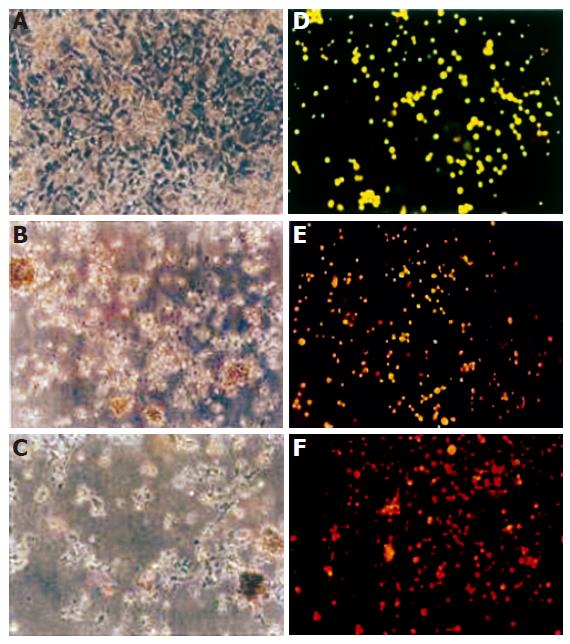
Figure 2 Morphological changes of HepG2 cells under light and fluorescence microscope.
HepG2 cells were incubated for 48 h with T. arjuna extract. The medium was removed and the cells were rinsed and visualized under light microscope in control (A), 60 mg/LL (B), 100 mg/L (C) of T. arjuna extract. Cells were stained with ethidium bromide and acridine orange and observed under fluorescence microscope in control (D), 60 mg/L (E) and 100 mg/L (F) of T. arjuna extract.
- Citation: Sivalokanathan S, Vijayababu MR, Balasubramanian MP. Effects of Terminalia arjuna bark extract on apoptosis of human hepatoma cell line HepG2. World J Gastroenterol 2006; 12(7): 1018-1024
- URL: https://www.wjgnet.com/1007-9327/full/v12/i7/1018.htm
- DOI: https://dx.doi.org/10.3748/wjg.v12.i7.1018


