Copyright
©2006 Baishideng Publishing Group Co.
World J Gastroenterol. Feb 21, 2006; 12(7): 1005-1012
Published online Feb 21, 2006. doi: 10.3748/wjg.v12.i7.1005
Published online Feb 21, 2006. doi: 10.3748/wjg.v12.i7.1005
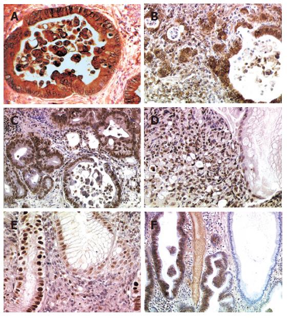
Figure 1 Immunohistochemical staining in human gastric adenocarcinoma and precancerous mucosa.
A: Intense cytoplasmic immunostaining of iNOS in tumor cells of tubular infiltrative carcinoma (TC), well differentiated; B: Intense cytoplasmic immunostaining of NTYR in tumor cells of TC, moderately differentiated; C: Intense nuclear immunostaining of 8-OH-dG in tumor cells of TC, well differentiated, note the reactivity of the lymphoid follicle (upper left); D: Intense nuclear immunostaining of 8-OH-dG in tumor cells of diffuse carcinoma, note the weak reactivity of some cells of a normal crypt (right); E: Intense nuclear immunostaining of 8-OH-dG in high-grade dysplastic epithelium, note the weak reactivity of a normal crypt (right); and F: Intense cytoplasmic immunostaining of NTYR in low-grade dysplastic epithelium, note the absence of reactivity of a normal crypt (right).
- Citation: Bancel B, Estève J, Souquet JC, Toyokuni S, Ohshima H, Pignatelli B. Differences in oxidative stress dependence between gastric adenocarcinoma subtypes. World J Gastroenterol 2006; 12(7): 1005-1012
- URL: https://www.wjgnet.com/1007-9327/full/v12/i7/1005.htm
- DOI: https://dx.doi.org/10.3748/wjg.v12.i7.1005


