Copyright
©2006 Baishideng Publishing Group Co.
World J Gastroenterol. Feb 14, 2006; 12(6): 987-989
Published online Feb 14, 2006. doi: 10.3748/wjg.v12.i6.987
Published online Feb 14, 2006. doi: 10.3748/wjg.v12.i6.987
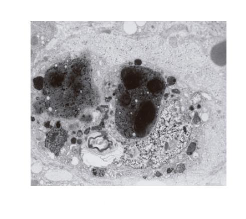
Figure 3 A hypertrophic, evidently activated Kupffer cell with phagocytized, very thick, black pigment deposits and biliary lamellar material.
The cell blocks the sinusoidal vascular lumen. Distinctly dilated Disse’s space contains a fine granular material (×4 400).
- Citation: Sobaniec-Lotowska ME, Lebensztejn DM. Ultrastructure of Kupffer cells and hepatocytes in the Dubin-Johnson syndrome: A case report. World J Gastroenterol 2006; 12(6): 987-989
- URL: https://www.wjgnet.com/1007-9327/full/v12/i6/987.htm
- DOI: https://dx.doi.org/10.3748/wjg.v12.i6.987


