Copyright
©2006 Baishideng Publishing Group Co.
World J Gastroenterol. Feb 14, 2006; 12(6): 987-989
Published online Feb 14, 2006. doi: 10.3748/wjg.v12.i6.987
Published online Feb 14, 2006. doi: 10.3748/wjg.v12.i6.987
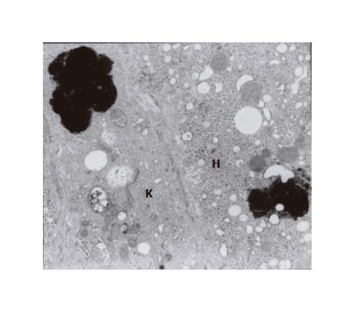
Figure 2 Fragment of Kupffer cell with phagocytized thick black pigment granules; the cell adheres to a hepatocyte (H) containing a similar pigment deposit (×7 000).
- Citation: Sobaniec-Lotowska ME, Lebensztejn DM. Ultrastructure of Kupffer cells and hepatocytes in the Dubin-Johnson syndrome: A case report. World J Gastroenterol 2006; 12(6): 987-989
- URL: https://www.wjgnet.com/1007-9327/full/v12/i6/987.htm
- DOI: https://dx.doi.org/10.3748/wjg.v12.i6.987


