Copyright
©2006 Baishideng Publishing Group Co.
World J Gastroenterol. Feb 14, 2006; 12(6): 982-986
Published online Feb 14, 2006. doi: 10.3748/wjg.v12.i6.982
Published online Feb 14, 2006. doi: 10.3748/wjg.v12.i6.982
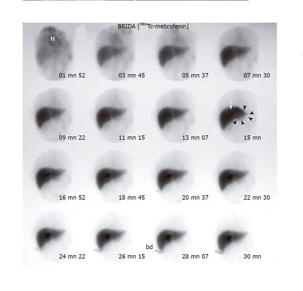
Figure 4 Postoperative 99mTc-mebrofenin dynamic study.
A well-circumscribed fusiform tracer accumulation in the right hepatic lobe near the hepatic hilum is clearly visible from the 15th min of the study (arrow), and corresponds to a dilated intrahepatic duct. A much fainter bulbous accumulation is apparent in the area of the left hepatic lobe, extending well beyond the liver margins (arrowheads) and corresponding to the extrahepatic portion of the choledochal cyst. H: heart blood pool; bd: biliary drainage.
- Citation: Stipsanelli E, Valsamaki P, Tsiouris S, Arka A, Papathanasiou G, Ptohis N, Lahanis S, Papantoniou V, Zerva C. Spontaneous rupture of a type IVA choledochal cyst in a young adult during radiological imaging. World J Gastroenterol 2006; 12(6): 982-986
- URL: https://www.wjgnet.com/1007-9327/full/v12/i6/982.htm
- DOI: https://dx.doi.org/10.3748/wjg.v12.i6.982


