Copyright
©2006 Baishideng Publishing Group Co.
World J Gastroenterol. Feb 14, 2006; 12(6): 982-986
Published online Feb 14, 2006. doi: 10.3748/wjg.v12.i6.982
Published online Feb 14, 2006. doi: 10.3748/wjg.v12.i6.982
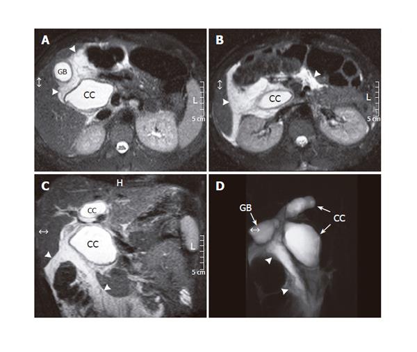
Figure 3 Abdominal MRI (spin echo T2-weighed, transverse projections - A and B, coronal projection - C) displaying bile leakage into the peritoneal cavity (arrowheads).
The study was followed by MRCP (turbo spin echo T2-weighed acquisition - D) which imaged the bile leak (arrowheads) and moreover revealed the “egg-timer” bicameral shape of the extrahepatic moiety of the choledochal cyst (CC). GB: gallbladder.
- Citation: Stipsanelli E, Valsamaki P, Tsiouris S, Arka A, Papathanasiou G, Ptohis N, Lahanis S, Papantoniou V, Zerva C. Spontaneous rupture of a type IVA choledochal cyst in a young adult during radiological imaging. World J Gastroenterol 2006; 12(6): 982-986
- URL: https://www.wjgnet.com/1007-9327/full/v12/i6/982.htm
- DOI: https://dx.doi.org/10.3748/wjg.v12.i6.982


