Copyright
©2006 Baishideng Publishing Group Co.
World J Gastroenterol. Feb 14, 2006; 12(6): 982-986
Published online Feb 14, 2006. doi: 10.3748/wjg.v12.i6.982
Published online Feb 14, 2006. doi: 10.3748/wjg.v12.i6.982
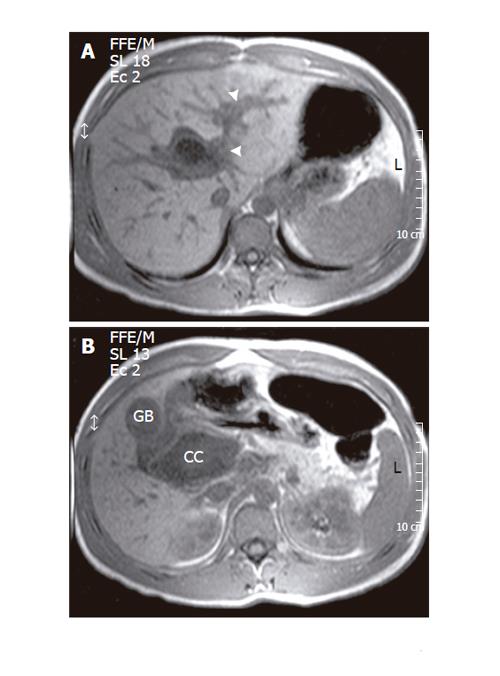
Figure 2 Significant distension of the intrahepatic bile ducts (arrowhead) in MRI of the upper abdomen (gradient echo T1-weighed acquisition) (A).
The walls of the choledochal cyst (CC) tend to acquire a concave shape, while its volume appears reduced as compared to the CT findings, both signs are considered as indicative of rupture (B). GB: gallbladder.
- Citation: Stipsanelli E, Valsamaki P, Tsiouris S, Arka A, Papathanasiou G, Ptohis N, Lahanis S, Papantoniou V, Zerva C. Spontaneous rupture of a type IVA choledochal cyst in a young adult during radiological imaging. World J Gastroenterol 2006; 12(6): 982-986
- URL: https://www.wjgnet.com/1007-9327/full/v12/i6/982.htm
- DOI: https://dx.doi.org/10.3748/wjg.v12.i6.982


