Copyright
©2006 Baishideng Publishing Group Co.
World J Gastroenterol. Feb 14, 2006; 12(6): 928-934
Published online Feb 14, 2006. doi: 10.3748/wjg.v12.i6.928
Published online Feb 14, 2006. doi: 10.3748/wjg.v12.i6.928
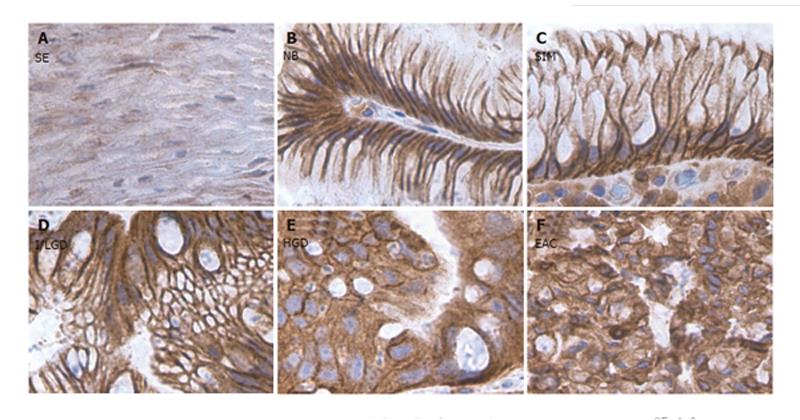
Figure 1 COX-2 staining in the sections from different stages of BE along with the sections from normal squamous and CLE nondetectable BE.
A: Squamous epithelium (SE); B: Nondetectable BE in CLE (NB); C: Specialized intestinal metaplasia (SIM); D: Indeterminate/low-grade dysplasia (I/LGD); E: high-grade dysplasia (HGD); F: Esophageal adenocarcinoma (EAC). Strong COX-2 staining was detected in both NCP cells and ACP cells in the histological stages of SIM, I/LGD, HGD and EAC. A weak COX-2 staining was observed in the normal squamous epithelium.
- Citation: Li Y, Wo JM, Ray MB, Jones W, Su RR, Ellis S, Martin RCG. Cyclooxygenase-2 and epithelial growth factor receptor up-regulation during progression of Barrett’s esophagus to adenocarcinoma. World J Gastroenterol 2006; 12(6): 928-934
- URL: https://www.wjgnet.com/1007-9327/full/v12/i6/928.htm
- DOI: https://dx.doi.org/10.3748/wjg.v12.i6.928


