Copyright
©2006 Baishideng Publishing Group Co.
World J Gastroenterol. Feb 14, 2006; 12(6): 921-927
Published online Feb 14, 2006. doi: 10.3748/wjg.v12.i6.921
Published online Feb 14, 2006. doi: 10.3748/wjg.v12.i6.921
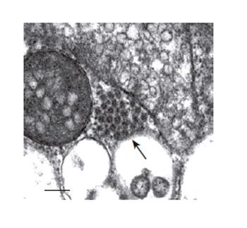
Figure 4 Electron microscopy examinations of EV71 VLP aggregates formed in cytoplasm of the Bac-P1-3CD-infected insect cells (100 000 X magnification).
The cells were infected at MOI 10, harvested at 3 dpi and ultrathin sectioned for TEM. The arrowhead indicates the VLP aggregate. Bar:100 nm.
- Citation: Chung YC, Huang JH, Lai CW, Sheng HC, Shih SR, Ho MS, Hu YC. Expression, purification and characterization of enterovirus-71 virus-like particles. World J Gastroenterol 2006; 12(6): 921-927
- URL: https://www.wjgnet.com/1007-9327/full/v12/i6/921.htm
- DOI: https://dx.doi.org/10.3748/wjg.v12.i6.921


