Copyright
©2006 Baishideng Publishing Group Co.
World J Gastroenterol. Feb 14, 2006; 12(6): 896-901
Published online Feb 14, 2006. doi: 10.3748/wjg.v12.i6.896
Published online Feb 14, 2006. doi: 10.3748/wjg.v12.i6.896
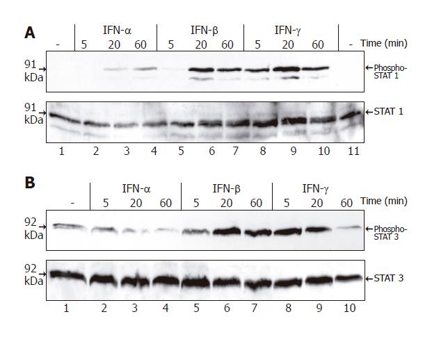
Figure 5 Tyrosine phosphorylation of STAT1 and STAT3 in PSCs induced by IFN.
PSCs growing in 6-well plates (one passage) were treated with IFN-α (106 U/L), IFN-β (106 U/L) and IFN-γ (100 μg/L) as indicated. Cell lysates were normalized for protein concentration and resolved by SDS-PAGE. A: Tyrosine phosphorylation of STAT1 assayed by immunoblotting using an antibody specific for the tyrosine-phosphorylated protein (upper panel). To control loading, the blot was stripped and reprobed with an anti-STAT1 protein-specific antibody (lower panel); B: Immunoblot analysis performed using an antibody specific for tyrosine-phosphorylated STAT3 (upper panel) and the blot stripped and reprobed with an anti-STAT3 protein-specific antibody (lower panel). Results are representative of three independent experiments.
- Citation: Baumert JT, Sparmann G, Emmrich J, Liebe S, Jaster R. Inhibitory effects of interferons on pancreatic stellate cell activation. World J Gastroenterol 2006; 12(6): 896-901
- URL: https://www.wjgnet.com/1007-9327/full/v12/i6/896.htm
- DOI: https://dx.doi.org/10.3748/wjg.v12.i6.896


