Copyright
©2006 Baishideng Publishing Group Co.
World J Gastroenterol. Feb 14, 2006; 12(6): 858-867
Published online Feb 14, 2006. doi: 10.3748/wjg.v12.i6.858
Published online Feb 14, 2006. doi: 10.3748/wjg.v12.i6.858
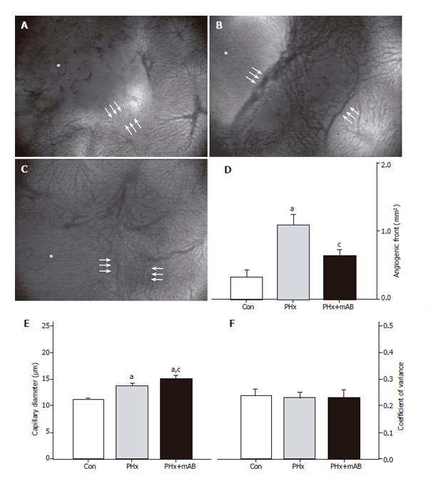
Figure 2 Analysis of the size of the angiogenic front (area between the triple arrows) at the margin of tumors (asterisks) in control mice (A, Con), after hepatectomy (B, PHx) and after hepatectomy and additional anti-MIP-2 treatment (C, PHx+mAB).
Quantitative analysis of the area of the angiogenic front (D) indicated a significant increase after hepatectomy (PHx) when compared with that of controls (Con). Additional anti-MIP-2 treatment (PHx+mAB) was capable of reducing the angiogenic process. Further, hepatectomy (PHx) significantly increased capillary dilation within the angiogenic front (E) compared to controls (Con), which was even more pronounced after additional anti-MIP-2 treatment (PHx+mAB). The heterogeneity of capillary diameters, as given by the coefficient of variance, was not affected in either of the treatment groups (F). Mean ± SE; aP < 0.05 vs Con; cP < 0.05 vs PHx. Magnifications (A-C) ×40.
- Citation: Kollmar O, Menger MD, Schilling MK. Macrophage inflammatory protein-2 contributes to liver resection-induced acceleration of hepatic metastatic tumor growth. World J Gastroenterol 2006; 12(6): 858-867
- URL: https://www.wjgnet.com/1007-9327/full/v12/i6/858.htm
- DOI: https://dx.doi.org/10.3748/wjg.v12.i6.858


