Copyright
©2006 Baishideng Publishing Group Co.
World J Gastroenterol. Feb 7, 2006; 12(5): 815-817
Published online Feb 7, 2006. doi: 10.3748/wjg.v12.i5.815
Published online Feb 7, 2006. doi: 10.3748/wjg.v12.i5.815
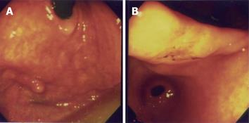
Figure 1 Esophagogastroduodenoscopic examination.
A: A 0.4-cm sessile polyp with a smooth surface is seen at the right upper posterior wall of the gastric fundus. B: An approximately 1.7 cm × 1.4 cm, depressed, flat white-red based lesion with oozing hemorrhages is present at the gastric angle.
- Citation: Lin YL, Tzeng JE, Wei CK, Lin CW. Small gastrointestinal stromal tumor concomitant with early gastric cancer: A case report. World J Gastroenterol 2006; 12(5): 815-817
- URL: https://www.wjgnet.com/1007-9327/full/v12/i5/815.htm
- DOI: https://dx.doi.org/10.3748/wjg.v12.i5.815


