Copyright
©2006 Baishideng Publishing Group Co.
World J Gastroenterol. Feb 7, 2006; 12(5): 812-814
Published online Feb 7, 2006. doi: 10.3748/wjg.v12.i5.812
Published online Feb 7, 2006. doi: 10.3748/wjg.v12.i5.812
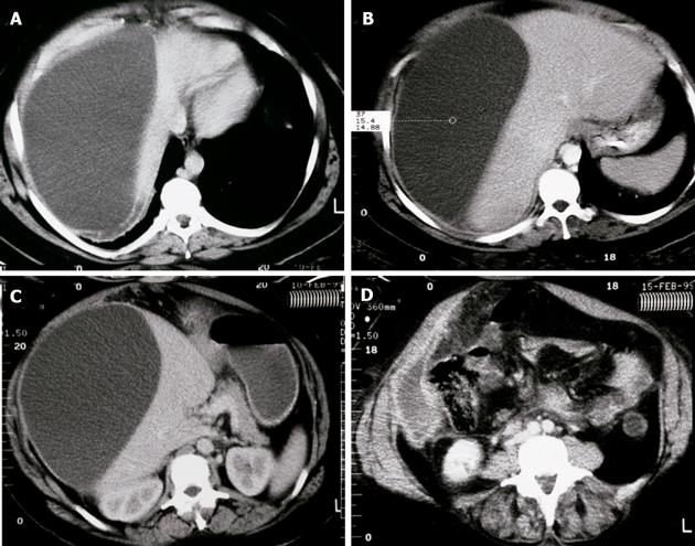
Figure 1 Abdominal CT scan.
A: Axial CT scan shows the large inhomo-geneous cystic fluid collection occupying the whole right lobe of the liver; B: CT scan of the cystic lesion on the level of spleen; C: the largest cystic lesion compresses the left lobe of the liver dislocating the nearby organs; D: cystic collection was seen also in the pararenal spaces and extended into the small pelvis.
- Citation: Battyány I, Herbert Z, Rostás T, Vincze &, Fülöp A, Harmat Z, Gasztonyi B. Successful percutaneous drainage of a giant hydatid cyst in the liver. World J Gastroenterol 2006; 12(5): 812-814
- URL: https://www.wjgnet.com/1007-9327/full/v12/i5/812.htm
- DOI: https://dx.doi.org/10.3748/wjg.v12.i5.812


