Copyright
©2006 Baishideng Publishing Group Co.
World J Gastroenterol. Feb 7, 2006; 12(5): 731-738
Published online Feb 7, 2006. doi: 10.3748/wjg.v12.i5.731
Published online Feb 7, 2006. doi: 10.3748/wjg.v12.i5.731
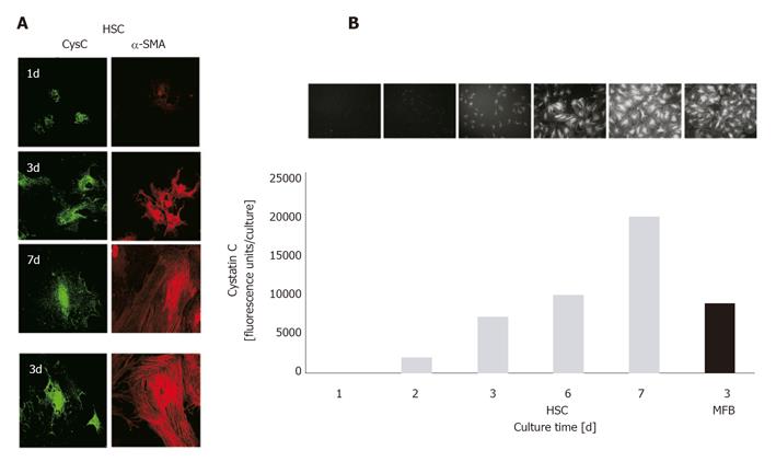
Figure 2 Confocal laser scanning microscopy and ELF assay for quantitative determination of CysC expression in HSCs and MFBs.
A: HSCs/MFBs were seeded on glass cover slides for indicated time intervals. After fixation, cells were permeabilized and incubated with primary antibodies directed against CysC or α-SMA. Primary antibodies were visualized with a FITC-labeled secondary antibody (CysC, green fluorescence) or a Cy3-labeled secondary antibody (α-SMA, red fluorescence). Original magnification was ×400. Negative controls, using normal IgGs instead of primary antibodies showed no staining (data not shown); B: HSCs/MFBs cultured in 96-well plates for indicated time intervals were fixed and stained for CysC. The staining was visualized by fluorescence microscopy (original magnification ×200). Fluorescence intensities were measured quantitatively using a 96-well fluorescence reader (excitation 365 nm, emission ≥515 nm).
- Citation: Gressner AM, Lahme B, Meurer SK, Gressner O, Weiskirchen R. Variable expression of cystatin C in cultured trans-differentiating rat hepatic stellate cells. World J Gastroenterol 2006; 12(5): 731-738
- URL: https://www.wjgnet.com/1007-9327/full/v12/i5/731.htm
- DOI: https://dx.doi.org/10.3748/wjg.v12.i5.731


