Copyright
©2006 Baishideng Publishing Group Co.
World J Gastroenterol. Dec 21, 2006; 12(47): 7649-7653
Published online Dec 21, 2006. doi: 10.3748/wjg.v12.i47.7649
Published online Dec 21, 2006. doi: 10.3748/wjg.v12.i47.7649
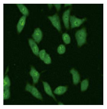
Figure 4 Presence of MK in the nucleoli of HepG2 cells.
The cells were stained with goat anti-MK antibody (1:1000). Cells were fixed with PFA and the secondary antibody FITC-conjugated mouse anti-goat antibody was added. The staining (green for MK) is presented (× 400).
- Citation: Dai LC, Xu DY, Yao X, Min LS, Zhao N, Xu BY, Xu ZP, Lu YL. Construction of a fusion protein expression vector MK-EGFP and its subcellular localization in different carcinoma cell lines. World J Gastroenterol 2006; 12(47): 7649-7653
- URL: https://www.wjgnet.com/1007-9327/full/v12/i47/7649.htm
- DOI: https://dx.doi.org/10.3748/wjg.v12.i47.7649


