Copyright
©2006 Baishideng Publishing Group Co.
World J Gastroenterol. Dec 21, 2006; 12(47): 7613-7620
Published online Dec 21, 2006. doi: 10.3748/wjg.v12.i47.7613
Published online Dec 21, 2006. doi: 10.3748/wjg.v12.i47.7613
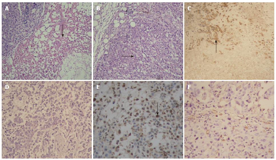
Figure 4 Pathological examination of tumor specimens (× 200).
HE staining showed wide areas of necrosis (arrow) in tumor tissues of the SG300-treated group (A), but cancer cells grew luxuriantly (arrow) in the control group (B). Immunohistochemistry demonstrated that most cancer cells around the necrotic area were positive for adenoviral capsid protein hexon (arrow) in the SG300-treated group (C), but cancer cells were negative for hexon in the control group (D). More cancer cells were positive for TUNEL labeling (arrow) in tumor tissues of the SG300-treated group (E), whereas only a few cancer cells were positive for TUNEL labeling in the control group (F).
- Citation: Su CQ, Wang XH, Chen J, Liu YJ, Wang WG, Li LF, Wu MC, Qian QJ. Antitumor activity of an hTERT promoter-regulated tumor-selective oncolytic adenovirus in human hepatocellular carcinoma. World J Gastroenterol 2006; 12(47): 7613-7620
- URL: https://www.wjgnet.com/1007-9327/full/v12/i47/7613.htm
- DOI: https://dx.doi.org/10.3748/wjg.v12.i47.7613


