Copyright
©2006 Baishideng Publishing Group Co.
World J Gastroenterol. Dec 14, 2006; 12(46): 7488-7496
Published online Dec 14, 2006. doi: 10.3748/wjg.v12.i46.7488
Published online Dec 14, 2006. doi: 10.3748/wjg.v12.i46.7488
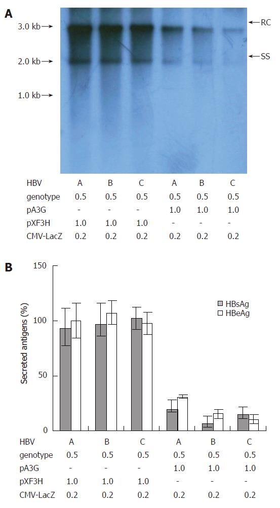
Figure 3 Suppression of replication of HBV clinical isolates of different genotypes by APOBEC3G.
(A) pHBV1.3 or the linear monomeric HBV genomes of genotype B and C were transiently co-transfected into HepG2 cells with the CMV-driven expression vector encoding A3G or empty vector pXF3H and pCMV-LacZ using Lipofectamine 2000 reagents. The cells were harvested 3 d after transfection. HBV core-associated viral DNA was prepared from nuclease-treated cytoplasmic lysates. Viral replicative DNA intermediates were analyzed by Southern blotting using a Dig-labeled full-length HBV DNA probe. (B) HBsAg and HBeAg levels were determined in the media of co-transfected HepG2 cells by ELISA, normalized to the activity of co-tansfected β-galactosidase in the cell lysates. The mean ± SEM of six independent experiments is shown (error bar indicates standard error). pA3G: APOBEC3G expression plasmids; RC: relaxed circular DNA; SS: single-stranded DNA.
- Citation: Lei YC, Tian YJ, Ding HH, Wang BJ, Yang Y, Hao YH, Zhao XP, Lu MJ, Gong FL, Yang DL. N-terminal and C-terminal cytosine deaminase domain of APOBEC3G inhibit hepatitis B virus replication. World J Gastroenterol 2006; 12(46): 7488-7496
- URL: https://www.wjgnet.com/1007-9327/full/v12/i46/7488.htm
- DOI: https://dx.doi.org/10.3748/wjg.v12.i46.7488


