Copyright
©2006 Baishideng Publishing Group Co.
World J Gastroenterol. Dec 7, 2006; 12(45): 7380-7387
Published online Dec 7, 2006. doi: 10.3748/wjg.v12.i45.7380
Published online Dec 7, 2006. doi: 10.3748/wjg.v12.i45.7380
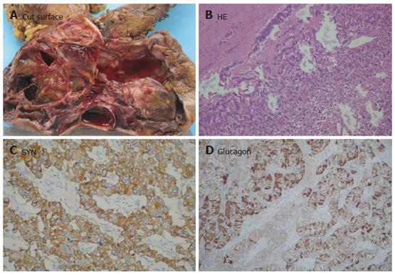
Figure 6 Cystic pancreatic endocrine tumor.
A: Cut surface of a large PET showing degeneration and cystic change; B: Tumor cells arranged as trabecular nests in the remnant area (HE × 100); C: SYN presented in all tumor cells (Envison × 200); D: Most tumor cells expressing glucagon in cytoplasm (Envison × 100).
- Citation: Ji Y, Lou WH, Jin DY, Kuang TT, Zeng MS, Tan YS, Zeng HY, Sujie A, Zhu XZ. A series of 64 cases of pancreatic cystic neoplasia from an institutional study of China. World J Gastroenterol 2006; 12(45): 7380-7387
- URL: https://www.wjgnet.com/1007-9327/full/v12/i45/7380.htm
- DOI: https://dx.doi.org/10.3748/wjg.v12.i45.7380


