Copyright
©2006 Baishideng Publishing Group Co.
World J Gastroenterol. Dec 7, 2006; 12(45): 7371-7374
Published online Dec 7, 2006. doi: 10.3748/wjg.v12.i45.7371
Published online Dec 7, 2006. doi: 10.3748/wjg.v12.i45.7371
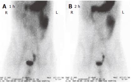
Figure 1 ECT examination showing small intestinal bleeding at 1 h (A) and 2 h (B) after intravenous injection of the tracer.
Serial planar imaging (1, 5, 15 and 30 min, 1 and 2 h) of the whole abdomen was performed. There was a radioactive accumulation at 5 min on the upper-left corner of the gall bladder, becoming increasingly dense till 2 h. Its emerging time was almost simultaneously as the image of the stomach with the density being slightly lower than that of the stomach image. During the whole period of imaging, its location was comparatively stable. The dynamic imaging of the whole abdomen showed an abnormal radioactive imaging near the upper-left corner of the gall bladder, which was considered the ectopic stomach mucous tissue inside the Meckel's diverticulum.
- Citation: Ba MC, Qing SH, Huang XC, Wen Y, Li GX, Yu J. Diagnosis and treatment of small intestinal bleeding: Retrospective analysis of 76 cases. World J Gastroenterol 2006; 12(45): 7371-7374
- URL: https://www.wjgnet.com/1007-9327/full/v12/i45/7371.htm
- DOI: https://dx.doi.org/10.3748/wjg.v12.i45.7371


