Copyright
©2006 Baishideng Publishing Group Co.
World J Gastroenterol. Dec 7, 2006; 12(45): 7292-7298
Published online Dec 7, 2006. doi: 10.3748/wjg.v12.i45.7292
Published online Dec 7, 2006. doi: 10.3748/wjg.v12.i45.7292
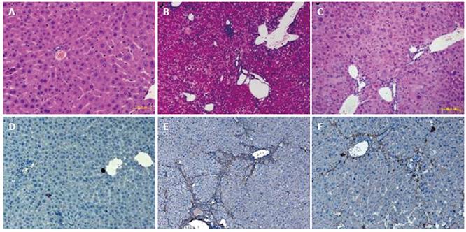
Figure 3 Effect of FLEP cell transplantation on liver fibrosis by HE staining and immunohistochemistry of α-SMA.
A: Normal liver tissue in control group (HE staining); B: Liver fibrosis in model group, fibrous tissue was formed with inflammatory cell infiltration in liver (HE staining); C: Liver fibrosis in FLEP cell transplantation group. The pathological change of liver was rather lighter compared with the model group (HE staining). D: Normal liver tissue, only α-SMA expression of vascular smooth muscle cell (immunohistochemical staining); E: Liver tissue in model group, displaying an increased amount of α-SMA expression with a dense network (immunohistochemical staining); F: Liver tissue in FLEP cell transplantation group, α-SMA expression was reduced compared with the model group (immunohistochemical staining). (original magnification, × 200).
- Citation: Zheng JF, Liang LJ, Wu CX, Chen JS, Zhang ZS. Transplantation of fetal liver epithelial progenitor cells ameliorates experimental liver fibrosis in mice. World J Gastroenterol 2006; 12(45): 7292-7298
- URL: https://www.wjgnet.com/1007-9327/full/v12/i45/7292.htm
- DOI: https://dx.doi.org/10.3748/wjg.v12.i45.7292


