Copyright
©2006 Baishideng Publishing Group Co.
World J Gastroenterol. Nov 28, 2006; 12(44): 7136-7142
Published online Nov 28, 2006. doi: 10.3748/wjg.v12.i44.7136
Published online Nov 28, 2006. doi: 10.3748/wjg.v12.i44.7136
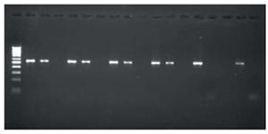
Figure 6 Typical gel image showing 16S rRNA amplification of H pylori.
Lane 1 represents 100bp Molecular weight marker, Lane 2 & 3, 5 & 6, 8 & 9, 11 & 12, 14 & 15 represents 16S rRNA amplification of H pylori DNA isolated from the culture. Lane 17 represents positive control of band at 534 bp. (ATCC26695), Lane 18 represents negative control.
-
Citation: Ibrahim M, Khan AA, Tiwari SK, Habeeb MA, Khaja M, Habibullah C. Antimicrobial activity of
Sapindus mukorossi andRheum emodi extracts againstH pylori :In vitro andin vivo studies. World J Gastroenterol 2006; 12(44): 7136-7142 - URL: https://www.wjgnet.com/1007-9327/full/v12/i44/7136.htm
- DOI: https://dx.doi.org/10.3748/wjg.v12.i44.7136


