Copyright
©2006 Baishideng Publishing Group Co.
World J Gastroenterol. Nov 21, 2006; 12(43): 6998-7006
Published online Nov 21, 2006. doi: 10.3748/wjg.v12.i43.6998
Published online Nov 21, 2006. doi: 10.3748/wjg.v12.i43.6998
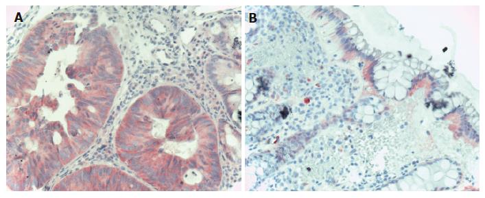
Figure 3 EGFR immunohistochemistry in CRC.
A: Carcinomatous glands of the colon showing diffuse cytoplasmatic, moderately intensive EGFR staining (Hematoxylin co-staining × 200); B: Normal colonic epithelia showing mildly, moderately intensive basolateral intracytoplasmatic EGFR staining. The lower 2/3 of the crypts do not show EGFR positivity (Hematoxylin co-staining × 100).
- Citation: Galamb O, Sipos F, Dinya E, Spisak S, Tulassay Z, Molnar B. mRNA expression, functional profiling and multivariate classification of colon biopsy specimen by cDNA overall glass microarray. World J Gastroenterol 2006; 12(43): 6998-7006
- URL: https://www.wjgnet.com/1007-9327/full/v12/i43/6998.htm
- DOI: https://dx.doi.org/10.3748/wjg.v12.i43.6998


