Copyright
©2006 Baishideng Publishing Group Co.
World J Gastroenterol. Nov 14, 2006; 12(42): 6818-6827
Published online Nov 14, 2006. doi: 10.3748/wjg.v12.i42.6818
Published online Nov 14, 2006. doi: 10.3748/wjg.v12.i42.6818
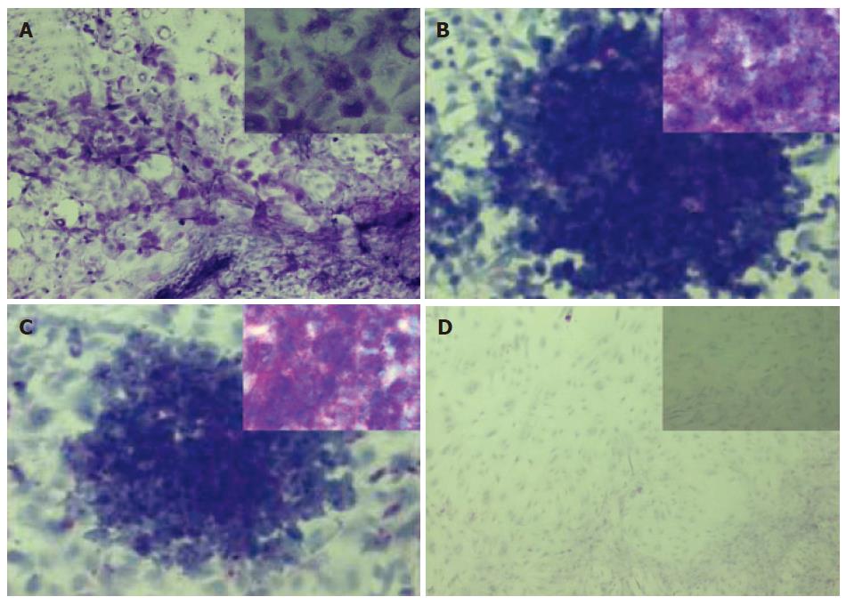
Figure 7 Histochemical staining of glycogen.
Periodic acid Schiff (PAS) staining was performed on cES cells co-cultured with MFLCs for 28 d (A) and undifferentiated cES cells (B), as well as those following treatment with α-amylase (C and D, respectively). Both the undifferentiated cES cells and those co-cultured with MFLCs showed violet staining (A, B). Violet staining was apparent in the undifferentiated cES cells (C), while such staining did not appear in the differentiated cES cells pretreated with α-amylase(D). Original magnification ×100 (A-D), × 400 (insets in A-D).
- Citation: Saito K, Yoshikawa M, Ouji Y, Moriya K, Nishiofuku M, Ueda S, Hayashi N, Ishizaka S, Fukui H. Promoted differentiation of cynomolgus monkey ES cells into hepatocyte-like cells by co-culture with mouse fetal liver-derived cells. World J Gastroenterol 2006; 12(42): 6818-6827
- URL: https://www.wjgnet.com/1007-9327/full/v12/i42/6818.htm
- DOI: https://dx.doi.org/10.3748/wjg.v12.i42.6818


