Copyright
©2006 Baishideng Publishing Group Co.
World J Gastroenterol. Nov 14, 2006; 12(42): 6818-6827
Published online Nov 14, 2006. doi: 10.3748/wjg.v12.i42.6818
Published online Nov 14, 2006. doi: 10.3748/wjg.v12.i42.6818
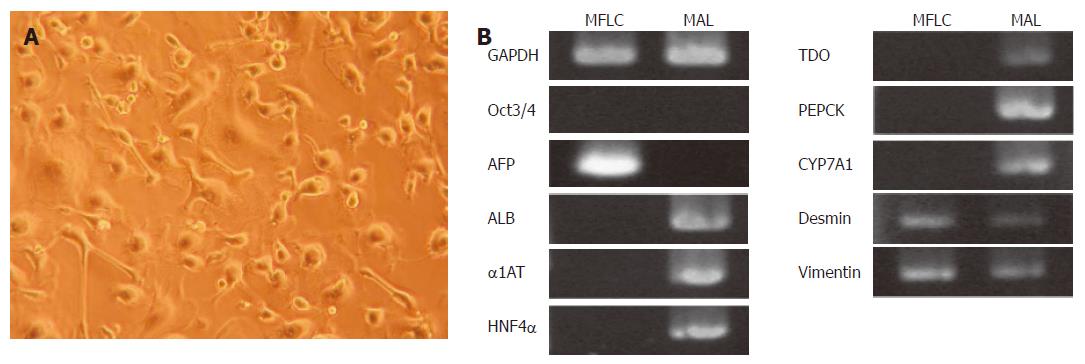
Figure 4 Characterization of MFLCs.
A: Optical microscope image of MFLCs (× 100). MFLCs were prepared from E14.0 mouse livers and cultured for 48 h. After the floating cell fraction was discarded from the culture, resting adherent cells were further cultured until semi-confluent. MFLCs showed various morphologies, including cuboidal and stellate-shaped cells; B: RT-PCR analysis of MFLCs. AFP, desmin, and vimentin were expressed, whereas ALB was not. MAL: Mouse adult liver cells.
- Citation: Saito K, Yoshikawa M, Ouji Y, Moriya K, Nishiofuku M, Ueda S, Hayashi N, Ishizaka S, Fukui H. Promoted differentiation of cynomolgus monkey ES cells into hepatocyte-like cells by co-culture with mouse fetal liver-derived cells. World J Gastroenterol 2006; 12(42): 6818-6827
- URL: https://www.wjgnet.com/1007-9327/full/v12/i42/6818.htm
- DOI: https://dx.doi.org/10.3748/wjg.v12.i42.6818


