Copyright
©2006 Baishideng Publishing Group Co.
World J Gastroenterol. Nov 14, 2006; 12(42): 6812-6817
Published online Nov 14, 2006. doi: 10.3748/wjg.v12.i42.6812
Published online Nov 14, 2006. doi: 10.3748/wjg.v12.i42.6812
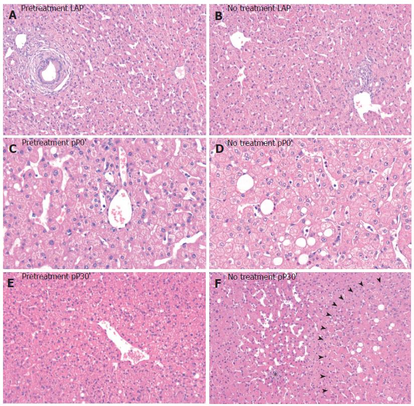
Figure 4 Representative liver sections after laparotomy (LAP), after 30 min of ischemia (pP0') and after 30 min of ischemia with 30 min of reperfusion (pP30') with and without 600 mg LA 15 min prior to ischemia, (HE×200-400).
A, B: In both groups no features of cell injury after LAP; C, D: At pP0' there was a mild oncotic injury in the untreated and pretreated group such as hepatocytes swelling and vacuolization; E, F: At pP30' oncotic injury was increased especially in the untreated group (F): there were areas with focal necrosis (asterisk) and areas with eosinophilia and oncosis (arrows). In the LA pretreated group oncotic cell injury was rare (E).
- Citation: Dünschede F, Erbes K, Kircher A, Westermann S, Seifert J, Schad A, Oliver K, Kiemer AK, Theodor J. Reduction of ischemia reperfusion injury after liver resection and hepatic inflow occlusion by α-lipoic acid in humans. World J Gastroenterol 2006; 12(42): 6812-6817
- URL: https://www.wjgnet.com/1007-9327/full/v12/i42/6812.htm
- DOI: https://dx.doi.org/10.3748/wjg.v12.i42.6812


