Copyright
©2006 Baishideng Publishing Group Co.
World J Gastroenterol. Nov 14, 2006; 12(42): 6797-6805
Published online Nov 14, 2006. doi: 10.3748/wjg.v12.i42.6797
Published online Nov 14, 2006. doi: 10.3748/wjg.v12.i42.6797
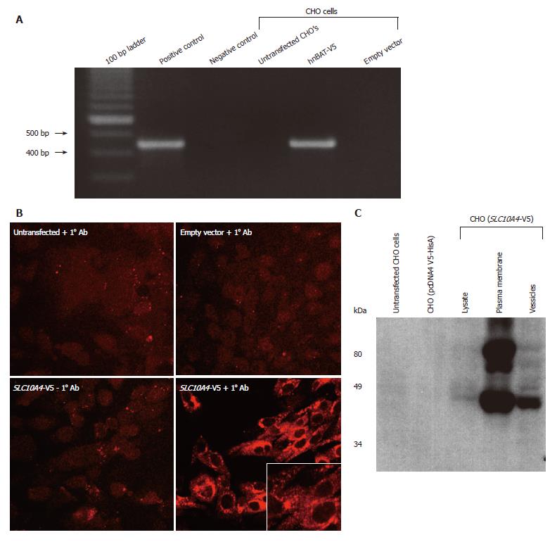
Figure 5 Analysis of the SLC10A4 stably transfected CHO cells.
(A) Expression of SLC10A4 mRNA in transfected CHO cells. SLC10A4 mRNA was detected by RT-PCR in the SLC10A4 transfected cells but not in untransfected CHO cells or the negative control (i.e., no template cDNA). (B) Protein expression of SLC10A4 in stably transfected CHO cells. No staining was found in untransfected CHO cells or CHO cells stably transfected with the empty vector (pcDNA4 V5-HisA) alone in the presence of the affinity-purified monoclonal V5 antibody, or in the CHO cells transfected with SLC10A4 in the absence of the primary antibody. Robust staining was detected in CHO cells transfected with SLC10A4 in the presence of the V5 antibody (× 40). (C) Immunoblotting analysis of SLC10A4 in transfected CHO cells. SLC10A4 is detected in the cell lysate (20 μg), mixed plasma membranes (40 μg) and vesicle (40 μg) fractions of the cells. No bands were seen in cell lysates (40 μg) from untransfected CHO cells or empty vector (pcDNA4 V5-HisA) stably transfected CHO cells.
- Citation: Splinter PL, Lazaridis KN, Dawson PA, LaRusso NF. Cloning and expression of SLC10A4, a putative organic anion transport protein. World J Gastroenterol 2006; 12(42): 6797-6805
- URL: https://www.wjgnet.com/1007-9327/full/v12/i42/6797.htm
- DOI: https://dx.doi.org/10.3748/wjg.v12.i42.6797


