Copyright
©2006 Baishideng Publishing Group Co.
World J Gastroenterol. Jan 28, 2006; 12(4): 652-655
Published online Jan 28, 2006. doi: 10.3748/wjg.v12.i4.652
Published online Jan 28, 2006. doi: 10.3748/wjg.v12.i4.652
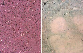
Figure 2 Macroscopic and microscopic examinations of tumor specimen.
A: Macroscopic examination showing well-defined non-encapsulated tumor. Microscopically, the tumor cells are arranged predominantly in a thin trabecular pattern. No portal triad can be seen in the tumor (HE×200); B: The non-neoplastic hepatocytes are almost normal, but contain cytoplasmic hemosiderin (Berlin Blue×20). On the other hand, no iron deposition is seen in the adenoma nodule.
- Citation: Hagiwara S, Takagi H, Kanda D, Sohara N, Kakizaki S, Katakai K, Yoshinaga T, Higuchi T, Nomoto K, Kuwano H, Mori M. Hepatic adenomatosis associated with hormone replacement therapy and hemosiderosis: A case report. World J Gastroenterol 2006; 12(4): 652-655
- URL: https://www.wjgnet.com/1007-9327/full/v12/i4/652.htm
- DOI: https://dx.doi.org/10.3748/wjg.v12.i4.652


