Copyright
©2006 Baishideng Publishing Group Co.
World J Gastroenterol. Oct 21, 2006; 12(39): 6397-6400
Published online Oct 21, 2006. doi: 10.3748/wjg.v12.i39.6397
Published online Oct 21, 2006. doi: 10.3748/wjg.v12.i39.6397
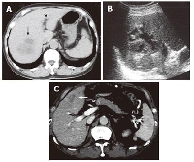
Figure 7 Computed tomography scan showing an abscess in the right anteroinferior segment of the liver (black arrow).
Pneumobilia is detected (black arrowhead) (A). Ultrasonography shows partial liquefaction in the abscess (B). Computed tomography scan showing an abscess in the left medial segment of the liver (white arrow) (C). Note that the pancreas is not swollen.
- Citation: Toshikuni N, Kai K, Sato S, Kitano M, Fujisawa M, Okushin H, Morii K, Takagi S, Takatani M, Morishita H, Uesaka K, Yuasa S. Pyogenic liver abscess after choledochoduodenostomy for biliary obstruction caused by autoimmune pancreatitis. World J Gastroenterol 2006; 12(39): 6397-6400
- URL: https://www.wjgnet.com/1007-9327/full/v12/i39/6397.htm
- DOI: https://dx.doi.org/10.3748/wjg.v12.i39.6397


