Copyright
©2006 Baishideng Publishing Group Co.
World J Gastroenterol. Oct 21, 2006; 12(39): 6316-6324
Published online Oct 21, 2006. doi: 10.3748/wjg.v12.i39.6316
Published online Oct 21, 2006. doi: 10.3748/wjg.v12.i39.6316
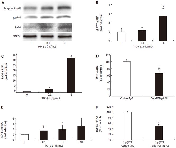
Figure 2 TGF-β1 stimulates the TGF-β signaling pathway.
A: The addition of 0.1 and 1 ng/mL TGF-β1 augmented Smad2 phosphorylation (phospho-Smad2), p15Ink4B, and PAI-1 proteins in Western blotting of HuCCT1 cells; B: TGF-β1 stimulation for 48 h enhanced p15Ink4B mRNA expression in HuCCT1 cells in RT-PCR: aP < 0.05 vs absence of TGF-β1; C: TGF-β1 stimulation for 48 h also augmented PAI-1 mRNA expression in HuCCT1 cells in RT-PCR: aP < 0.05 vs absence of TGF-β1; D: PAI-1 mRNA was determined by RT-PCR after incubating the HuCCT1 cells with 5 ng/mL neutralizing anti-TGF-β1 antibody or nonimmune control IgG for 96 h. The addition of anti-TGF-β1 antibody attenuated the level of PAI-1 mRNA: aP < 0.05 vs control IgG; E: The addition of TGF-β1 to HuCCT1 for 6 h led to a concentration-dependent increase in TGF-β1 mRNA expression by RT-PCR: aP < 0.05 vs absence of TGF-β1; F: TGF-β1 mRNA in HuCCT1 was determined by RT-PCR after incubating the cells with 5 ng/mL neutralizing anti-TGF-β1 antibody or nonimmune control IgG for 96 h. The addition of anti-TGF-β1 antibody attenuated the level of TGF-β1 mRNA: aP < 0.05 vs control IgG.
- Citation: Shimizu T, Yokomuro S, Mizuguchi Y, Kawahigashi Y, Arima Y, Taniai N, Mamada Y, Yoshida H, Akimaru K, Tajiri T. Effect of transforming growth factor-β1 on human intrahepatic cholangiocarcinoma cell growth. World J Gastroenterol 2006; 12(39): 6316-6324
- URL: https://www.wjgnet.com/1007-9327/full/v12/i39/6316.htm
- DOI: https://dx.doi.org/10.3748/wjg.v12.i39.6316


