Copyright
©2006 Baishideng Publishing Group Co.
World J Gastroenterol. Oct 21, 2006; 12(39): 6316-6324
Published online Oct 21, 2006. doi: 10.3748/wjg.v12.i39.6316
Published online Oct 21, 2006. doi: 10.3748/wjg.v12.i39.6316
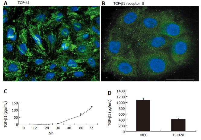
Figure 1 Expression of TGF-β1 and TGF-β receptor II in intrahepatic cholangiocarcinoma cell lines.
Immunofluorescence showed staining of TGF-β1 (A) and TGF-β receptor II (B) in HuCCT1 cells. Scale bar = 20 μm. C: Detection of TGF-β1 in supernatant of HuCCT1 by ELISA. TGF-β1 levels in the supernatant of HuCCT1 showed a time-dependent increase; D: The other ICC cell lines were incubated for 48 h and assayed by ELISA to determine the levels of TGF-β1 in the supernatants. MEC and HuH-28 cells also secreted TGF-β1 into the supernatants.
- Citation: Shimizu T, Yokomuro S, Mizuguchi Y, Kawahigashi Y, Arima Y, Taniai N, Mamada Y, Yoshida H, Akimaru K, Tajiri T. Effect of transforming growth factor-β1 on human intrahepatic cholangiocarcinoma cell growth. World J Gastroenterol 2006; 12(39): 6316-6324
- URL: https://www.wjgnet.com/1007-9327/full/v12/i39/6316.htm
- DOI: https://dx.doi.org/10.3748/wjg.v12.i39.6316


