Copyright
©2006 Baishideng Publishing Group Co.
World J Gastroenterol. Oct 14, 2006; 12(38): 6219-6224
Published online Oct 14, 2006. doi: 10.3748/wjg.v12.i38.6219
Published online Oct 14, 2006. doi: 10.3748/wjg.v12.i38.6219
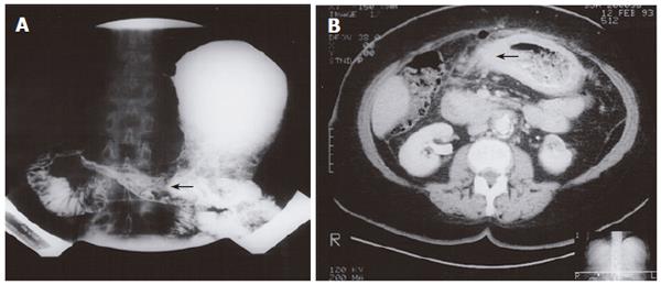
Figure 4 Double-contrast upper GI examination demonstrating irregular narrowing of the gastric antrum (arrow) (A), CT demonstrating gastric mural thickening, perigastric stranding, (arrow) and lymphadenopathy (B).
- Citation: Nazareno J, Taves D, Preiksaitis HG. Metastatic breast cancer to the gastrointestinal tract: A case series and review of the literature. World J Gastroenterol 2006; 12(38): 6219-6224
- URL: https://www.wjgnet.com/1007-9327/full/v12/i38/6219.htm
- DOI: https://dx.doi.org/10.3748/wjg.v12.i38.6219


