Copyright
©2006 Baishideng Publishing Group Co.
World J Gastroenterol. Oct 14, 2006; 12(38): 6149-6155
Published online Oct 14, 2006. doi: 10.3748/wjg.v12.i38.6149
Published online Oct 14, 2006. doi: 10.3748/wjg.v12.i38.6149
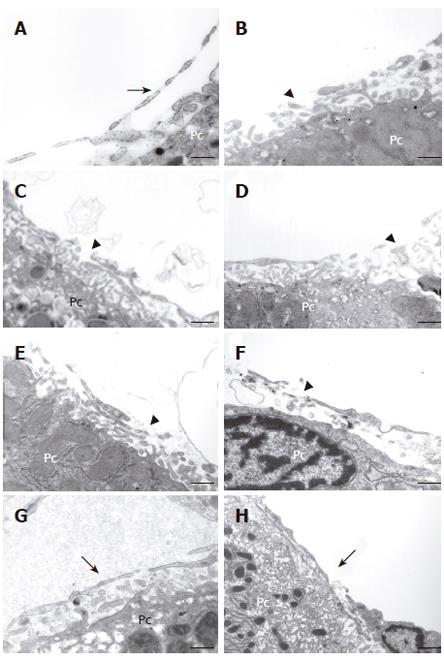
Figure 2 High magnification electron micrographs of liver sinusoids following hydrodynamic injection.
A: Control image illustrates intact histological relationship between liver sinusoidal endothelial and neighboring liver parenchymal cells (Pc). Note the fenestrated processes (arrow) of endothelial cells; B: Ten minutes after hydrodynamic injections, severe damage of the endothelial lining in the form of gaps is noted (arrowhead). Similar features were observed at 1 (C), 8 (D), 24 (E), and 72 (F) h, i.e, a disrupted endothelial lining with the presence of large structural rearrangement in the form of gaps (arrowheads). The architecture of hepatocytes remained intact when compared to the control. Between seven (G) to ten (H) days post injection, the number of gaps decreased significantly. Interestingly, the hepatic sinusoidal endothelial lining is less fenestrated when compared to the control (arrow). Scale bars, 1 μm.
- Citation: Yeikilis R, Gal S, Kopeiko N, Paizi M, Pines M, Braet F, Spira G. Hydrodynamics based transfection in normal and fibrotic rats. World J Gastroenterol 2006; 12(38): 6149-6155
- URL: https://www.wjgnet.com/1007-9327/full/v12/i38/6149.htm
- DOI: https://dx.doi.org/10.3748/wjg.v12.i38.6149


