Copyright
©2006 Baishideng Publishing Group Co.
World J Gastroenterol. Oct 14, 2006; 12(38): 6133-6141
Published online Oct 14, 2006. doi: 10.3748/wjg.v12.i38.6133
Published online Oct 14, 2006. doi: 10.3748/wjg.v12.i38.6133
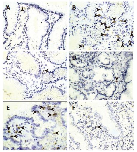
Figure 6 Apoptosis-related proteins in the gastric mucosa stained with immunoperoxidase.
Photomicrographs show the antral mucosa stained for FasL in a non-infected control patient (A), at the ulcer margin (B), and after eradication therapy of the same patient (C). Immunostaining for perforin is shown in a patient with H pylori-associated gastritis (D), and in a patient with gastric ulcer before (E), and after eradication therapy (F), respectively. Arrowheads show immunoreactive cells (Original magnification × 400).
-
Citation: Souza HS, Neves MS, Elia CC, Tortori CJ, Dines I, Martinusso CA, Madi K, Andrade L, Castelo-Branco MT. Distinct patterns of mucosal apoptosis in
H pylori -associated gastric ulcer are associated with altered FasL and perforin cytotoxic pathways. World J Gastroenterol 2006; 12(38): 6133-6141 - URL: https://www.wjgnet.com/1007-9327/full/v12/i38/6133.htm
- DOI: https://dx.doi.org/10.3748/wjg.v12.i38.6133


