Copyright
©2006 Baishideng Publishing Group Co.
World J Gastroenterol. Oct 7, 2006; 12(37): 6077-6079
Published online Oct 7, 2006. doi: 10.3748/wjg.v12.i37.6077
Published online Oct 7, 2006. doi: 10.3748/wjg.v12.i37.6077
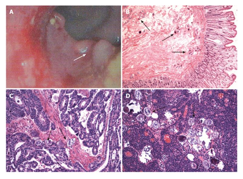
Figure 1 Colonoscopy showing an exophytic fragile neoplasm with an ulcerating surface (as indicated by the white arrow head) in the sigmoid colon (A), pathological analysis revealing deposited S.
japonicum ova and granuloma formation as well as fibrotic deposition in the submucosa of the sigmoid colon (B) (HE × 50), moderately- differentiated tubular adenocarcinoma (HE × 200) and deposited S. japonicum ova in the tumor (C), and deposited S. japonicum ova in the mesenteric lymph node (D) (HE × 200). Black arrow heads indicate S. japonicum ova.
- Citation: Li WC, Pan ZG, Sun YH. Sigmoid colonic carcinoma associated with deposited ova of Schistosoma japonicum: A case report. World J Gastroenterol 2006; 12(37): 6077-6079
- URL: https://www.wjgnet.com/1007-9327/full/v12/i37/6077.htm
- DOI: https://dx.doi.org/10.3748/wjg.v12.i37.6077


