Copyright
©2006 Baishideng Publishing Group Co.
World J Gastroenterol. Sep 28, 2006; 12(36): 5904-5906
Published online Sep 28, 2006. doi: 10.3748/wjg.v12.i36.5904
Published online Sep 28, 2006. doi: 10.3748/wjg.v12.i36.5904
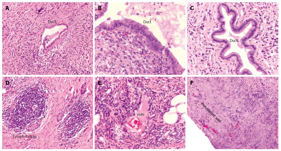
Figure 3 Histological findings of the resected pancreatic specimen.
Histological examination displaying marked chronic periductal lymphoplasmacytic inflammation and fibrosis (A-C), intrapancreatic lymph follicle (D), and venulitis (E). F depicts the wall of the pseudocyst without evidence of epithelial lining.
- Citation: Welsch T, Kleeff J, Esposito I, Büchler MW, Friess H. Autoimmune pancreatitis associated with a large pancreatic pseudocyst. World J Gastroenterol 2006; 12(36): 5904-5906
- URL: https://www.wjgnet.com/1007-9327/full/v12/i36/5904.htm
- DOI: https://dx.doi.org/10.3748/wjg.v12.i36.5904


