Copyright
©2006 Baishideng Publishing Group Co.
World J Gastroenterol. Sep 7, 2006; 12(33): 5412-5415
Published online Sep 7, 2006. doi: 10.3748/wjg.v12.i33.5412
Published online Sep 7, 2006. doi: 10.3748/wjg.v12.i33.5412
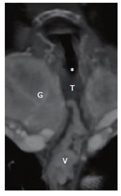
Figure 2 3D reconstruction of CT angiography with view at the dorsal wall of the trachea (T) demonstrating a venous plexus of downhill varices (V) on the wall between the esophagus and trachea connected with a thyroid vein at the goiter (G) of the right thyroid lobe; *, out of plane, cut off level thick slice.
- Citation: Veldt AAVD, Hadithi M, Paul MA, Berg FGVD, Mulder CJ, Craanen ME. An unusual cause of hematemesis: Goiter. World J Gastroenterol 2006; 12(33): 5412-5415
- URL: https://www.wjgnet.com/1007-9327/full/v12/i33/5412.htm
- DOI: https://dx.doi.org/10.3748/wjg.v12.i33.5412


