Copyright
©2006 Baishideng Publishing Group Co.
World J Gastroenterol. Sep 7, 2006; 12(33): 5393-5395
Published online Sep 7, 2006. doi: 10.3748/wjg.v12.i33.5393
Published online Sep 7, 2006. doi: 10.3748/wjg.v12.i33.5393
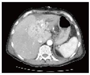
Figure 1 CT scan shows Klatskin tu-mor type III b with intrahepatic bile duct enlargement, cholesta-sis and atrophy of the left hepatic lobe.
- Citation: Schmeding M, Neumann U, Neuhaus P. Colonic metastasis of Klatskin tumor: Case report and discussion of the current literature. World J Gastroenterol 2006; 12(33): 5393-5395
- URL: https://www.wjgnet.com/1007-9327/full/v12/i33/5393.htm
- DOI: https://dx.doi.org/10.3748/wjg.v12.i33.5393


