Copyright
©2006 Baishideng Publishing Group Co.
World J Gastroenterol. Sep 7, 2006; 12(33): 5320-5325
Published online Sep 7, 2006. doi: 10.3748/wjg.v12.i33.5320
Published online Sep 7, 2006. doi: 10.3748/wjg.v12.i33.5320
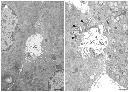
Figure 6 Transmission electron micrographs of isolated hepatocytes.
A bar indicates 1 μm. A: Normal hepatocytes prior to the addition of taurolithocholate (TLC). A bile canaliculus (BC) was slightly dilated and filled with microvilli. B: 12 h after the addition of 50 μmol/L TLC. Note the characteristic lamellar transformation of the bile canalicular membranes. A bile canaliculus (BC) was deformed and dilated with a loss of microvilli. Electron dense membranous structures of biliary materials (arrowheads) were found in the vacuoles in the pericanalicular region.
- Citation: Watanabe N, Kagawa T, Kojima SI, Takashimizu S, Nagata N, Nishizaki Y, Mine T. Taurolithocholate impairs bile canalicular motility and canalicular bile secretion in isolated rat hepatocyte couplets. World J Gastroenterol 2006; 12(33): 5320-5325
- URL: https://www.wjgnet.com/1007-9327/full/v12/i33/5320.htm
- DOI: https://dx.doi.org/10.3748/wjg.v12.i33.5320


