Copyright
©2006 Baishideng Publishing Group Co.
World J Gastroenterol. Sep 7, 2006; 12(33): 5320-5325
Published online Sep 7, 2006. doi: 10.3748/wjg.v12.i33.5320
Published online Sep 7, 2006. doi: 10.3748/wjg.v12.i33.5320
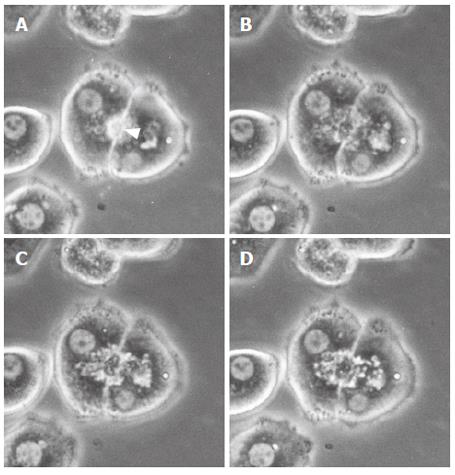
Figure 2 Representative phase contrast micrographs of an isolated hepatocyte couplet in the taurolithocholate (TLC)-treated groups (50 μmol/L).
Prior to addition (A) and after the addition of TLC (B, C, D). A canaliculus (arrowhead) between two hepatocytes (A) was deformed, and canalicular bile was incorporated into the vacuoles in the hepatocyte cytoplasm immediately after the addition of TLC (B). Thereafter, the bile canaliculi were gradually dilated, and the canalicular contractions were markedly impaired (C, D). In the TLC-treated groups, the movements of small particles became extremely slow, and cytoplasmic vesicles and vacuoles were noted in the pericanalicular region.
- Citation: Watanabe N, Kagawa T, Kojima SI, Takashimizu S, Nagata N, Nishizaki Y, Mine T. Taurolithocholate impairs bile canalicular motility and canalicular bile secretion in isolated rat hepatocyte couplets. World J Gastroenterol 2006; 12(33): 5320-5325
- URL: https://www.wjgnet.com/1007-9327/full/v12/i33/5320.htm
- DOI: https://dx.doi.org/10.3748/wjg.v12.i33.5320


