Copyright
©2006 Baishideng Publishing Group Co.
World J Gastroenterol. Aug 28, 2006; 12(32): 5229-5233
Published online Aug 28, 2006. doi: 10.3748/wjg.v12.i32.5229
Published online Aug 28, 2006. doi: 10.3748/wjg.v12.i32.5229
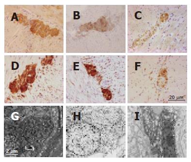
Figure 2 Representative examples illustrating the general neuronal marker NSE (A, B, C) and BCL-2 (D, E, F) immunoreactivities in the neuromuscular layer of a control subject (A, D), in the non dilated loop distal to the congenital obstruction of the patient (B, E) and in the dilated loop proximal to the congenital obstruction of the patient (C, F).
Note the marked reduction of NSE and BCL-2 immunoreactivities in myenteric ganglion cells and nerve fibers targeting the muscular layer observed only in the dilated loop. Streptavidin biotin immunoperoxidase technique. Calibration bar (A-F): 20 μm. Compared to controls (G) and the non-dilated segment (H), myenteric neurons of the present case (representative example in I) showed chromatin clumping, cell body shrinkage and cytoplasmic vacuolization. Uranyl acetate and lead citrate staining, transmission electron microscopy. Calibration bar (G, H, I): 2 μm.
- Citation: Nardo GD, Stanghellini V, Cucchiara S, Barbara G, Pasquinelli G, Santini D, Felicani C, Grazi G, Pinna AD, Cogliandro R, Cremon C, Gori A, Corinaldesi R, Sanders KM, Giorgio RD. Enteric neuropathology of congenital intestinal obstruction: A case report. World J Gastroenterol 2006; 12(32): 5229-5233
- URL: https://www.wjgnet.com/1007-9327/full/v12/i32/5229.htm
- DOI: https://dx.doi.org/10.3748/wjg.v12.i32.5229


