Copyright
©2006 Baishideng Publishing Group Co.
World J Gastroenterol. Aug 28, 2006; 12(32): 5175-5181
Published online Aug 28, 2006. doi: 10.3748/wjg.v12.i32.5175
Published online Aug 28, 2006. doi: 10.3748/wjg.v12.i32.5175
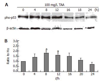
Figure 5 A: Phospho-p53 protein level (typical data, Western blot); B: Alterations of phospho-p53 protein as compared to H0 following β-actin normalization of clone 9 after treatment with 100 mg/L TAA for various times.
Data are presented as mean ± SE. aP < 0.05 vs the control (0 h). Individual experiments were repeated three times and each time point of treatment was triplicate.
- Citation: Chen LH, Hsu CY, Weng CF. Involvement of P53 and Bax/Bad triggering apoptosis in thioacetamide-induced hepatic epithelial cells. World J Gastroenterol 2006; 12(32): 5175-5181
- URL: https://www.wjgnet.com/1007-9327/full/v12/i32/5175.htm
- DOI: https://dx.doi.org/10.3748/wjg.v12.i32.5175


