Copyright
©2006 Baishideng Publishing Group Co.
World J Gastroenterol. Aug 28, 2006; 12(32): 5175-5181
Published online Aug 28, 2006. doi: 10.3748/wjg.v12.i32.5175
Published online Aug 28, 2006. doi: 10.3748/wjg.v12.i32.5175
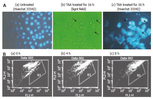
Figure 4 A: Morphology of clone 9 cell; (a) untreated (Hoechst 33342 staining); (b) after treatment with 100 mg/L TAA treatment for 16 h (light field); (c) after treatment with 100 mg/L mmol/L TAA for 16 h (Hoechst 33342, 5 mg/mL in PBS).
The arrow indicates the apoptotic cells; B: Flow cytometry of apoptosis in clone 9 cells (TUNEL assay) after treatment with 100 mg/L TAA for various times (a) 0 (b) 4 and (c) 8 h. R1 presents the area of cell apoptosis. Individual experiments were repeated three times and each time point of treatment was triplicate.
- Citation: Chen LH, Hsu CY, Weng CF. Involvement of P53 and Bax/Bad triggering apoptosis in thioacetamide-induced hepatic epithelial cells. World J Gastroenterol 2006; 12(32): 5175-5181
- URL: https://www.wjgnet.com/1007-9327/full/v12/i32/5175.htm
- DOI: https://dx.doi.org/10.3748/wjg.v12.i32.5175


