Copyright
©2006 Baishideng Publishing Group Co.
World J Gastroenterol. Aug 21, 2006; 12(31): 5071-5074
Published online Aug 21, 2006. doi: 10.3748/wjg.v12.i31.5071
Published online Aug 21, 2006. doi: 10.3748/wjg.v12.i31.5071
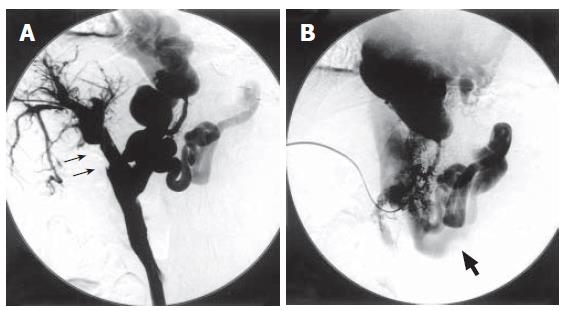
Figure 3 Percutaneous transhepatic portography after catheter-directed thrombolysis.
A: Dissolution of the thrombus in portal vein (narrow arrow); B: Hepatofugal flow into dilated portosystemic shunts (thick arrow: left renal vein).
- Citation: Nakai M, Sato M, Sahara S, Kawai N, Kimura M, Maeda Y, Ibata Y, Higashi K. Transhepatic catheter-directed thrombolysis for portal vein thrombosis after partial splenic embolization in combination with balloon-occluded retrograde transvenous obliteration of splenorenal shunt. World J Gastroenterol 2006; 12(31): 5071-5074
- URL: https://www.wjgnet.com/1007-9327/full/v12/i31/5071.htm
- DOI: https://dx.doi.org/10.3748/wjg.v12.i31.5071


