Copyright
©2006 Baishideng Publishing Group Co.
World J Gastroenterol. Aug 21, 2006; 12(31): 5071-5074
Published online Aug 21, 2006. doi: 10.3748/wjg.v12.i31.5071
Published online Aug 21, 2006. doi: 10.3748/wjg.v12.i31.5071
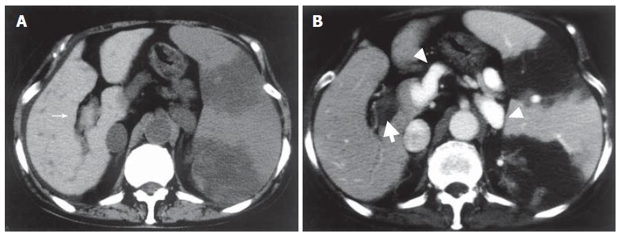
Figure 1 Abdominal CT one week after PSE.
A: Plain CT showing high density lesions in the main portal vein and the 1st right branch (narrow arrow); B: Contrast-enhanced CT revealing no enhancement of portal vein indicating portal thrombosis (thick arrow) and portosystemic shunts (arrow head). Splenic infarction after PSE was also visualized.
- Citation: Nakai M, Sato M, Sahara S, Kawai N, Kimura M, Maeda Y, Ibata Y, Higashi K. Transhepatic catheter-directed thrombolysis for portal vein thrombosis after partial splenic embolization in combination with balloon-occluded retrograde transvenous obliteration of splenorenal shunt. World J Gastroenterol 2006; 12(31): 5071-5074
- URL: https://www.wjgnet.com/1007-9327/full/v12/i31/5071.htm
- DOI: https://dx.doi.org/10.3748/wjg.v12.i31.5071


